Musculoskeletal (MSK) imaging radiologists focus on diagnosing bone and joint abnormalities, including orthopedic, rheumatologic and traumatic conditions. By specializing in musculoskeletal imaging, these physician experts are capable of focusing on additional nuances related to orthopedic and sports medicine injuries.
MSK imaging subspecialists work in partnership with primary care physicians, surgeons, sports medicine, orthopedic specialists and more. They interpret a variety of examinations depending on a patient’s needs.
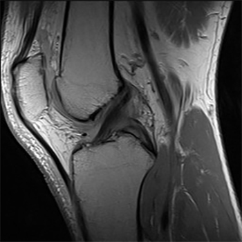
MRI | Magnetic resonance imaging (MRI) uses a powerful magnetic field, radio waves and a computer to produce detailed pictures of the body’s internal structures that are clearer, more detailed and more likely in some instances to identify and accurately characterize disease than other imaging methods.
Ultrasound | Ultrasound imaging uses a transducer or probe to generate sound waves and produce pictures of the body’s internal structures. It does not use ionizing radiation, has no known harmful effects, and provides a clear picture of soft tissues that don’t show up well on x-ray images.
X-Ray & Fluoroscopy | X-ray uses a very small dose of ionizing radiation to produce pictures of the body’s internal structures. X-rays are the oldest and most frequently used form of medical imaging.
To learn more about these procedures visit www.radiologyinfo.org