Neurological imaging subspecialists (neuroradiologists) use a variety of techniques to obtain images of the head, neck and spine. This information is used to diagnose and treat a variety of conditions such as stroke, Alzheimer’s disease, cancer, headaches, hearing loss, multiple sclerosis and brain tumors.
Neuroradiologists work closely with neurologists, primary care providers, surgeons, neurointerventional specialists and others to provide a coordinated, informed and effective approach to neurological patient care. Neurological imaging procedures include MRI, PET, CT and ultrasound.
CTA | CT angiography (CTA) uses a CT scanner to produce detailed images of both blood vessels and tissues in various parts of the body. An iodine-rich contrast material (dye) is usually injected through a small catheter placed in a vein of the arm. A CT scan is then performed while the contrast flows through the blood vessels to the various organs of the body.
MRA | Magnetic resonance angiography (MRA) uses a powerful magnetic field, radio frequency waves and a computer to produce detailed images of the major arteries within the body, evaluate blood vessels and help identify any abnormalities. It does not use ionizing radiation (x-rays) and can be performed with or without contrast material.
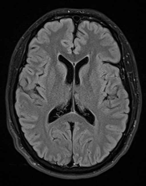
MRI | Magnetic resonance imaging (MRI) uses a powerful magnetic field, radio waves and a computer to produce detailed pictures of the body’s internal structures that are clearer, more detailed and more likely in some instances to identify and accurately characterize disease than other imaging methods.
Myelography | Myelography uses a real-time form of x-ray called fluoroscopy and an injection of contrast material to evaluate the spinal cord, nerve roots and spinal lining (meninges). It is particularly useful for assessing the spine following surgery and for assessing disc abnormalities in patients who cannot undergo MRI.
PET/CT | Combines the metabolic activity results of a PET Scan with the precise anatomical localization of a CT Scan. This allows our physicians to pinpoint the exact location of cancerous tumors within the body.
Ultrasound | Ultrasound imaging uses a transducer or probe to generate sound waves and produce pictures of the body’s internal structures. It does not use ionizing radiation, has no known harmful effects, and provides a clear picture of soft tissues that don’t show up well on x-ray images.
To learn more about these procedures visit www.radiologyinfo.org