Breast imaging radiologists are clinical experts who work as a team to use best practices for early detection of breast cancer, providing compassionate care and optimal outcomes. They must meet strict criteria for quality and adhere to the highest standards of care.
Breast imaging subspecialists work closely with breast surgeons, oncologists, radiation oncologists, OB/GYNs, family practitioners and other health care professionals. They utilize a variety of procedures, depending on a patient’s needs to develop a comprehensive treatment plan. Breast imaging examinations/procedures include mammography, 3D mammography, ultrasound, MRI, breast biopsy and breast needle localization.
Breast Ultrasound | This exam allows physicians to better characterize breast lesions, such as whether they are solid or fluid-filled.
Breast Specific Gamma Imaging (BSGI) or Scintimammography | First, a radiotracer, or drug that emits radioactivity, is injected. Then, because the radiotracer accumulates differently in different kinds of tissue, it can help physicians determine whether cancer could be present, thus helping determine whether a biopsy or additional follow-up is necessary.
Mammography (Screening & Diagnostic) | A screening mammogram is an annual exam performed as a preventative measure. It is recommended that all women over the age of 40 receive an annual screening mammogram. A diagnostic mammogram is used to look more closely at a specific area of abnormality.
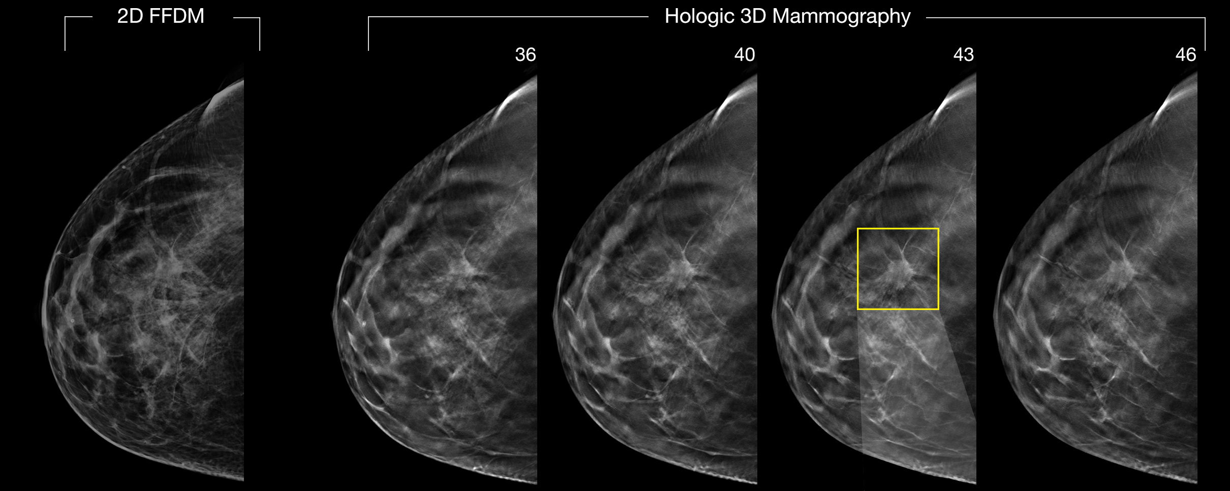
3D (Digital Breast Tomography or DBT) Mammography | 3D Mammography creates a three dimensional picture of the breast using X-rays. It represents a huge leap forward in detecting breast cancer in its earliest stages.
3D Mammographic Guided Breast Biopsy | 3D Mammographic biopsy utilizes 3D imaging to help find the abnormality and remove a tissue sample for further microscopic evaluation. It’s less invasive than a surgical biopsy and leaves little to no scarring.
Ultrasound Guided Breast Biopsy | An ultrasound-guided breast biopsy is a procedure that uses sound waves to first localize a lump or abnormality and then remove a tissue sample for examination under a microscope. It is less invasive than surgical biopsy, leaves little to no scarring and does not involve exposure to ionizing radiation.
MRI Guided Breast Biopsy | An MRI breast biopsy is performed when a breast lump or abnormality is only visible on MRI. It is less invasive than a surgical biopsy, leaves little or no scarring and does not involve exposure to ionizing radiation.
Steorotactic Guided Breast Biopsy | This procedure uses mammography – a specific type of breast imaging that uses low-dose x-rays – to help locate a breast lump or abnormality and remove a tissue sample for examination under a microscope. It is less invasive than surgical biopsy, leaves little to no scarring and can be an excellent way to evaluate calcium deposits or tiny masses that are not visible on ultrasound.
PET/CT | This exam combines the metabolic activity results of a PET Scan with the precise anatomical localization of a CT Scan. This allows our physicians to pinpoint the exact location of cancerous tumors within the body.
To learn more about these procedures visit www.radiologyinfo.org
Catawba Radiology’s local data shows that nearly 20% of the cancers detected by a screening mammogram were in women ages 40-49, further supporting the start of screening mammograms at the age of 40. Additionally 40% of the life years lost to breast cancer are in women diagnosed in their 40s.