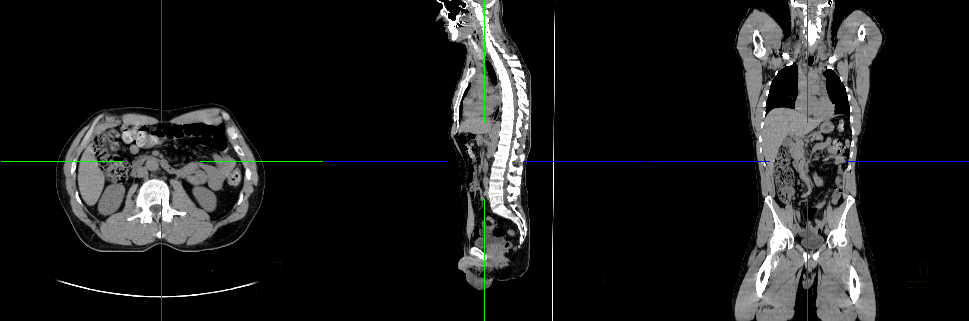
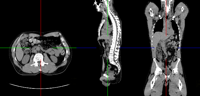
Body imaging specialists diagnose and help treat diseases and conditions (particularly cancer) affecting major body organs and systems, including the lungs, liver, stomach, spine, pelvis, kidneys, colon, pancreas, as well as other structures. A subspecialized body imaging radiologist conducts imaging exams from the throat down to the pelvis. These radiologists use many types of imaging, including MRI, CT, Ultrasound and PET/CT to see inside the body in ways never before imaginable.
Body imagers also perform minimally invasive procedures from biopsies and drainages to radiofrequency ablations of tumors to spare the patient from having surgery.
CT Lung Cancer Screening | Screening examinations are tests performed to find disease before symptoms begin. The goal of screening is to detect disease at its earliest and most treatable stage. Lung cancer is cancer that forms in tissues of the lung, usually in the cells lining air passages. Lung cancer that is detected early — before spreading to other areas of the body — is more successfully treated. In lung cancer screening, individuals who have a high risk of developing lung cancer but no signs or symptoms of the disease undergo low-dose computed tomography (LDCT) scanning of the chest.
MRI | Magnetic resonance imaging (MRI) uses a powerful magnetic field, radio waves and a computer to produce detailed pictures of the body’s internal structures that are clearer, more detailed and more likely in some instances to identify and accurately characterize disease than other imaging methods.
Needle Biopsy – Lung Nodule | Needle biopsy of the lung uses imaging guidance to help locate a nodule or abnormality and remove a tissue sample for examination under a microscope. A biopsy may be necessary when imaging tests cannot confirm that a nodule is benign, or a nodule cannot be reached by bronchoscopy or other methods.
PET/CT | Combines the metabolic activity results of a PET Scan with the precise anatomical localization of a CT Scan. This allows our physicians to pinpoint the exact location of cancerous tumors within the body.
Ultrasound | Ultrasound imaging uses a transducer or probe to generate sound waves and produce pictures of the body’s internal structures. It does not use ionizing radiation, has no known harmful effects, and provides a clear picture of soft tissues that don’t show up well on x-ray images.
CT Colonography | Less invasive exam that uses low dose radiation CT scanning to obtain an interior view of the colon to examine the large intestine for cancer and growths called polyps.
X-Ray & Fluoroscopy | X-ray or radiography uses a very small dose of ionizing radiation to produce pictures of the body’s internal structures. X-rays are the oldest and most frequently used form of medical imaging.
To learn more about these procedures visit www.radiologyinfo.org